
|
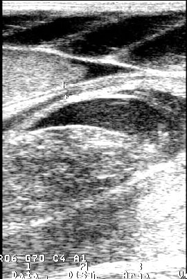
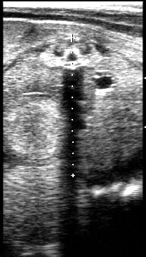
This image is obtained with the transducer in a sagittal position with respect to the uterus on the left side of the stingray. The large and small + symbols denote the uterine wall thickness (0.6 cm). The mucosal surface forms villous extensions or trophonemata which allow the uterus to supply nutrients to the fetus. Histotroph or uterine milk is secreted by trohponemata crypts and delivered to the fetus (Hamlett et al., 2005).
This particular stingray produced trophonemata with or without an egg capsule but viable fetuses were never seen. It is speculated that optimal conditions for pregnancy were not met for this particular animal. |
|
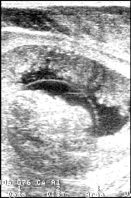
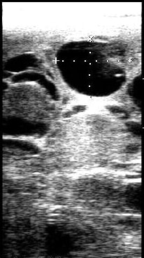
This image is obtained with the transducer in a sagittal position with respect to the uterus on the left side of the stingray.
The large and small + symbols denote the uterine wall thickness (0.3 cm) during this early stage of gestation. A distinct capsule is seen which is an egg capsule in this species. After ovulation an egg capsule may form. If copulation occurs then fertilization may occur as well or the sperm may be stored until optimum conditions are identified. This particular stingray was examined once every other week for four months. The egg capsule remained and at approximately the same size. The liver length decreased over this time period from over 11 cm to 4 cm. |
|
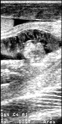
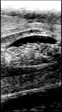
This image is obtained with the transducer in a transverse position with respect to the uterus on the left side of the stingray.
This image shows the uterus with developed trophonemata and egg capsule. This egg capsule is fertilized and this is during early gestation. This particular stingray is examined here five months prior to parturition. |
|
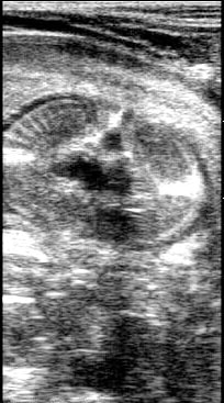
This image is obtained with the transducer in a sagittal position with respect to the uterus on the left side of the stingray.
This image shows the uterus with distinct trophonemata within the uterus. The round structure within the uterus (yellow overlay) is suspected to be an egg yolk sac which helps provide nutrients to the embryo during development. The caudal tip of the liver is also seen here. This particular stingray was monitored for only two more months then moved and fetal development was not followed. |
|
This image is obtained with the transducer in a transverse position with respect to the fetus.
This ultrasound image shows the cranial aspect of the fetus. The dark center of the fetus is the oral cavity to the esophagus which will narrow and widen as the stingray moves fluid through the gills and to the stomach. The dorsal aspect of the pup shows another anechoic area surrounded by a hyperechoic area which is the cartilaginous spine. This particular stingray pupped this litter two months later. |
|
This image is obtained with the transducer in a transverse position relative to the fetus on the left side of the pregnant stingray.
This image shows the caudal aspect of the fetus (coelomic cavity of the fetus). Within the uterine wall the fetus occupies the majority of the space. The large and small + symbols along with the dotted line denote the overall thickness of the fetus (3.92 cm). The cartilage forming the spine at the dorsal aspect of the fetus produces an acoustic shadow. The gall bladder and spiral intestine are also distinctly seen here. This particular stingray pupped one month after this exam was conducted. |
|
This image is obtained with the transducer in a sagittal position with respect to the uterus on the left side of the stingray.
This image shows the uterus approximately one week after the stingray pupped. The uterus and its contents are shown as well as the liver. This particular stingray gave birth to two full size pups and one mummified fetus. |
|
This image is obtained with the transducer in a sagittal position with respect to the ovaries on the left side of the stingray.
This image shows the epigonal organ embedded with multiple cystic ovaries. The large and small x and + symbols denote the dimensions of the cystic ovary (2.08 cm x 1.65 cm). This image is from the same stingray whose uterus was shown under Post-gestation. The uterus during this examination was enlarged and fluid-filled. This examination was performed approximately 18 months later. This condition is possibly a reproductive disease that occurs when housed without males and experiencing elevated estradiol and low progestin (sezarc.org) |
All images were captured using a commmercial ultrasound unit (Aloka SSD-900v) and 7.5 MHz linear array transducer. The overall gain, time gain compensation, and depth settings were adjusted in order to maximize image resolution and organ visualization.
